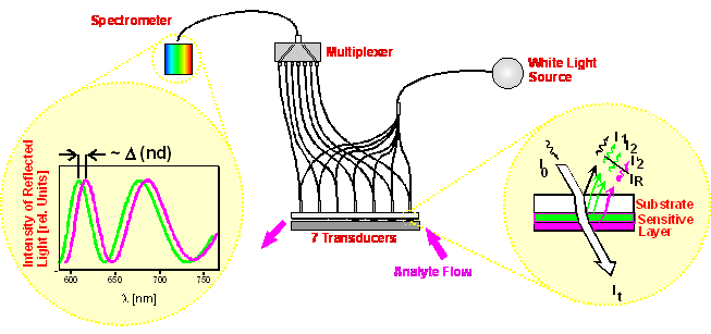
The Reflectometric
Interference Spectroscopy (RIfS), which was initially proposed by Gauglitz and
Nahm [175],
evaluates the interference pattern of light, which is reflected at thin transparent
films. The interference is caused by different partial beams, which are reflected
in a different way at the film interfaces depending on the wavelength. Changes
of the optical thickness nd of
the sensitive layer can be detected as shifts of the maxima and minima of the
interference pattern [176].
The RIfS
sensor array shown in figure 12 contains 7 sensors
allowing the application of 7 sensitive layers. The configuration of the sensor
array is based on a white light source. The light is transmitted through a lens
and filter system via polymer fibers to the glass substrates with 7 different
polymer layers. The reflected light is transmitted to an optical multiplexer,
which is directly connected to a diode array spectrometer. More details can
be found elsewhere [177].
figure 12: Schematic of the RIfS
array setup. The yellow bubbles show the effects when analyte sorbs into the
polymer layer (green before sorption of analyte, magenta after sorption of analyte).